Microstructure Imaging Core

Westin
Carl-Fredrik Westin, PhD
Core PI


Our Publications

The goal of the Microstructure Imaging Core is to pursue technological developments that improve current understanding of white matter anatomy and pathology in the brain. Given the small dimension of the neural pathways in relation to current imaging resolution, these technologies are vital to analyzing and visualizing the neural anatomy in normal and diseased states. Our current clinical objective is to analyze abnormalities in white matter architecture that underlie schizophrenia by using techniques in diffusion magnetic resonance imaging (dMRI) and post-process imaging. The expectation is that new knowledge acquired from these studies not only will benefit the understanding and treatment of schizophrenia, but also a broad range of other white matter diseases. The work of this core is organized around the following themes:

  1. Development of Microstructural Imaging Biomarkers
  2. Development of Personalized Anatomical Analysis Methods
  3. Population Statistics and Patient-Specific Diagnosis

The objective of this research is to develop and implement quantitative measures from diffusion MRI (dMRI). Methods to be explored include multi-shell dMRI to determine free vs. multi-compartmental or restricted diffusion; compressed sensing (CS) in conjunction with MRS to allow detection of microstructural metabolic change within the tissue; and double-pulsed field gradient dMRI (double-PFG) to provide previously unavailable diffusion properties that can be mapped into families of new types of geometric and tissue-specific parameters. Software for measuring the proposed biomarkers will be developed and tested.

Featured Technologies

Multishell DMRI 
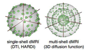
Standard dMRI (left) allows measurement of the angular distribution of the diffusion function; whereas the radial part of this function measured with multi-shell dMRI (right) reflects the type of diffusion observed (free vs. multicompartmental, or restricted).


Double PFG 
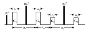
Traditional diffusion MRI (single-PFG) would only provide the first pair of gradients, G1,G1. Increased microstructural detail (G2, G2) can be measured with the double-PFG diffusion sequence.

Diffusion MRI in 3D Slicer

Double PFG

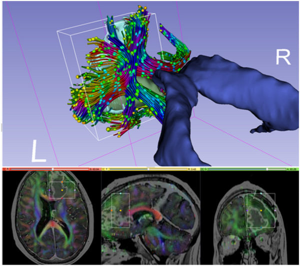
Image legend: Diffusion MRI data for neurosurgical planning. The tractography region of interest (ROI) is depicted by the box placed around the tumor (in green) in the frontal lobe. The ROI is also visualized with rectangles in the slice views below. Tracts are then created based on the principal diffusion directions, which are color coded (bottom). Diffusion ellipsoids are shown along the tract to visualize the shape of the local diffusion.

The 3D Slicer (or simply Slicer) software was initially developed as a joint effort by the Surgical Planning Laboratory of Brigham and Women's Hospital and the Medical Vision Group of the MIT-based Computer Science and Artificial Intelligence Laboratory (CSAIL). The goal was to create an interactive, open source software platform for biomedical research. The program has evolved into a national platform supported by a variety of federal funding sources. This versatile research environment has resulted in a wide array of functionality and supports a variety of medical imaging projects.

Slicer is a "point and click" end-user application. It is used as a vehicle for delivering algorithms to computer scientists, biomedical researchers, and clinical investigators. Slicer is distributed under an open source license without a reciprocity requirement and without restrictions on use. For a sampling of the portfolio of applications, please see the Slicer Community page. 3D Slicer consists of over a million lines of code, mostly C++. This massive software development effort was enabled by the participation of several large-scale NIH-funded efforts, including the National Alliance for Medical Image Computing (NA-MIC), Neuroimage Analysis Center (NAC), Biomedical Informatics Research Network (BIRN), Center for Integration of Medicine and Innovative Technology (CIMIT), and National Center for Image Guided Therapy (NCIGT) communities. Federal funding sources include the National Center for Research Resources (NCRR), National Institute of Biomedical Imaging and Bioengineering (NIBIB), NIH Roadmap, National Cancer Institute (NCI), National Science Foundation (NSF), and the Department of Defense (DOD) among others.

DTMRI is a module in the 3D Slicer collection used for interactive visualization of diffusion tensor MRI (DT-MRI, or DTI). A cross-platform end user application used for analyzing and visualizing medical images, Slicer consists of a collection of Open Source libraries for developing and deploying new image computing technologies. It is an algorithm development platform with a powerful new Execution Model to facilitate creation of new modules. The White Matter Architecture Core has made major contributions in the diffusion MRI analysis and visualization packages in Slicer2, Slicer3, and Slicer4 (www.slicer.org).

Estimation Procedures for High Angular Resolution Diffusion MRI

OPTD Method

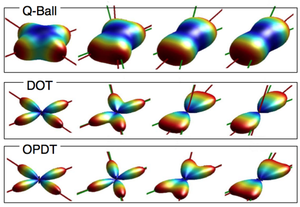
The White Matter Architure Core has developed novel estimation procedures for High Angular Resolution Diffusion MRI (HARDI). Our novel method “Orientation Probability Density Transform” (OPDT) of estimating the diffusion function has received widespread attention [Tristan-Vega, 2009], [Tristan-Vega, 2010]. Compared to the popular approaches of Q-Ball imaging and the diffusion orientation transform (DOT), our formulation introduces considerably less blurring of the angular function as shown in the figure.

In our work, instead of radial projections, true angular marginalizations were computed. We showed that the Jacobian of the spherical coordinates (in this case r2) needs to be included, a crucial contribution as the transform has been calculated without the correct term since 2003. Our formulation, the OPDT, is advantageous as it has a strict probabilistic interpretation, and the correct expression introduces considerably less blurring of the angular function in comparison to other methods such as Q-Ball and DOT.

The figure shows diffusion profiles for crossing fibers with different angles reconstructed using the Q-Ball imaging (top), the diffusion orientation transform (middle), and our formulation (OPDT). Note that the OPTD method introduces considerably less blurring of the angular function, making it easier to detect the peaks of the function corresponding to the two underlying diffusion directions.
Image legend: Diffusion profiles for crossing fibers with different angles reconstructed using the Q-Ball imaging (top), the diffusion orientation transform (middle), and our formulation (OPDT).

Multiple Fiber Model Tractography

Unscented Kalman Filtering

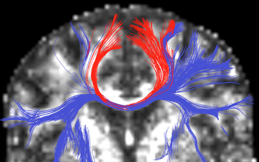
Image legend: Multiple fiber model tractography using unscented Kalman filtering. Note the additional tracts (blue) the new method finds.

The White Matter Architecture Core has developed tractography methods that can trace complex white matter regions, such as regions where there is heavy crossing and branching of the fiber tracts [Malcolm, 2009]. In this work we propose a technique to simultaneously estimate the local fiber orientations and perform multi-fiber tractography. Existing techniques estimate the local fiber orientation at each voxel independently. Consequently, there is no running knowledge of confidence in the measured signal or estimated fiber orientation. Further, to overcome noise, many algorithms use a filter as a post-processing step to obtain a smooth trajectory. Fiber tracking is formulated as a causal estimation: at each step of tracing the fiber, the current estimate of the signal is guided by the previous. The signal is modeled as a discrete mixture of Watson directional functions, and the tractography is performed within a filtering framework. Starting from a seed point, each fiber is traced to its termination using an unscented Kalman filter to simultaneously fit the signal and propagate in the most consistent direction. Despite the presence of noise and uncertainty, this provides an accurate estimate of the local structure at each point along the fiber. The Watson function was chosen since it provides a compact representation of the signal parameterized by the principal diffusion direction and a scaling parameter describing anisotropy. It also allows analytic reconstruction of the oriented diffusion function from those parameters. Using a mixture of two and three components (corresponding to two-fiber and three-fiber models) we demonstrate in synthetic experiments that this approach reduces signal reconstruction error and significantly improves the angular resolution at crossings and branchings. In vivo experiments shown in the figure examine the corpus callosum and internal capsule and confirm the ability to trace through regions known to contain such crossing and branching while providing inherent path regularization.

Tract-Based Diffusion Feature Analysis

EM Slicer Interface

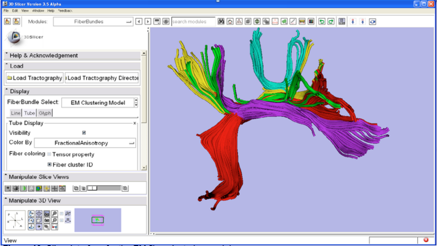
Image legend: SLICER interface for the EM fiber clustering module.

The White Matter Architecture Core has also worked on developing an automatic method that we call tract-based morphometry, or TBM, for measurement and analysis of diffusion MRI data along white matter fiber tracts [O'Donnell, 2009]. Using subject-specific tractography bundle segmentations, the method generates an arc length parameterization of the bundle with point correspondences across all fibers and all subjects, allowing tract-based measurement and analysis. In this work the team presents a quantitative comparison of fiber coordinate systems from the literature and we introduce an improved optimal match method that reduces spatial distortion and improves intra- and inter-subject variability of FA measurements.

The team proposed a method for generating arc length correspondences across hemispheres, enabling a TBM study of interhemispheric diffusion asymmetries in the arcuate fasciculus (AF) and cingulum bundle (CB). The results of this study demonstrate that TBM can detect differences that may not be found by measuring means of scalar invariants in entire tracts, such as the mean diffusivity (MD) differences found in AF. TBM results are reported of higher fractional anisotropy (FA) in the left hemisphere in AF (caused primarily by lower λ3, the smallest eigenvalue of the diffusion tensor, in the left AF), and higher left hemisphere FA in CB (related to higher λ1, the largest eigenvalue of the diffusion tensor, in the left CB). By mapping the significance levels onto the tractography trajectories for each structure, the anatomical locations of the interhemispheric differences are shown. The TBM approach brings analysis of DTI data into the clinically and neuroanatomically relevant framework of the tract anatomy. The team has also developed tools for tract-based analysis in 3D Slicer. As shown in the figure, the new command line module for EM clustering developed in collaboration with GE Research (Jim Miller) makes anatomical grouping and quantitative analysis of diffusion measures available to the research community. The team has also created an on-line tutorial for this module.
 

Asymmetry of White Matter Tract

Odonnell Handedness

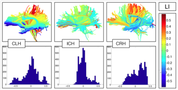
Image legend: Our white matter clustering method defines homologous anatomy across subjects (left). It has been validated by comparison to interactive tract selection (middle). The clustering method has been used in several studies of schizophrenia (SZ), e.g., to define callosal subdivisions, detecting reduced FA in the anterior corpus callosum in SZ (right).

The team has proposed a new method for studying the asymmetry of white matter tracts in the entire brain. This approach has been applied to a preliminary study of normal subjects across the handedness spectrum [O’Donnell, 2010]. The quantification of brain asymmetries may provide biomarkers for presurgical localization of language function and can improve our understanding of neural structure-function relationships in health and disease. Methods for quantifying white matter asymmetry using diffusion MRI tractography have thus far been based on comparing numbers of fibers or volumes of a single fiber tract across hemispheres. The team further proposes a generalization of such methods, where the “number of fibers” laterality measurement is extended to the entire brain using a soft fiber comparison metric. The distribution of fiber laterality indices over the whole brain is summarized in a histogram, and defines properties of the distribution using its skewness, median, and inter-quartile range. The whole-brain fiber laterality histogram can be measured in an exploratory fashion without hypothesizing asymmetries only in particular structures. The team has demonstrated an overall difference in white matter asymmetry in consistent- and inconsistent-handers: the skewness of the fiber laterality histogram is significantly different across handedness groups. The figure shows fiber laterality indices and fiber laterality histograms in example subjects from each handedness group.

Research Highlights

CF1
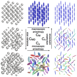
Figure 1: Q-space trajectory imaging for multidimensional diffusion MRI of the human brain. This visualization of four proposed QTI measures demonstrates how the measures would change in eight illustrative synthetic macrodomains (voxels). Note that these measures intuitively separate size, shape, and orientation coherence, as well as provide the traditional measures of macroscopic anisotropy. The microstructure measures proposed from QTI can disambiguate complex microenvironments that are indistinguishable using today’s standard single diffusion encoding (SDE) methods. For example, the macroscopic anisotropy (FA) of all diffusion tensor distributions (DTDs) found in the bottom row is very low, while the microscopic anisotropy (μFA) reflects the anisotropy of the individual micro-environments [Westin, 2016].

CF2

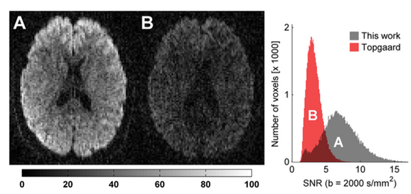
Figure 2: Constrained optimization of gradient waveforms for generalized diffusion encoding. The team provided a solution to the problem of constrained optimization of gradient waveforms imposed by clinical hardware, while maximizing the diffusion encoding strength afforded by QTI, by solving a constrained optimization problem that accommodates constraints on maximum gradient amplitude, slew rate, coil heating and positioning of radio frequency pulses. The method's efficacy and flexibility is demonstrated both experimentally and by comparison with previous work on optimization of isotropic diffusion sequences. In this figure, raw diffusion weighted images are shown on an axial slice through the corpus callosum in a healthy volunteer. The encoding strength is b=2000 s/mm2 in both images. The measured signal is markedly higher in the images encoded with the optimized waveform (A, echo time 116 ms) compared to the qMAS waveform (B, echo time 170 ms). The histogram shows the distribution of voxelwise SNR from brain tissue located within the imaging slab. There is a clear tendency towards higher SNR for the optimized waveform, due to the shorter echo time [Sjölund, 2015].

CF3

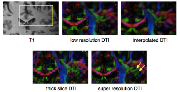
Figure 3A: Sampling strategies and compressed sensing for high spatial resolution dMRI. T1 weighted MRI image with a yellow inset showing the corresponding inset of the colored-by-orientation DTI images. The low resolution images have 1.83 mm3 isotropic voxels, while the interpolated DTI and the super resolution data set have 0.93 mm3 isotropic voxels. The yellow arrows point out the fine details in the super resolution results that are missing in the low resolution and the interpolated results.

CF4

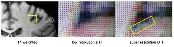
Figure 3B: Sampling strategies and compressed sensing for high spatial resolution dMRI. B. Left: T1 weighted MRI images with a yellow inset indicating the anatomical area of the DTI images with color-coded tensor glyphs visualizing the local white matter fiber orientation. Middle: the low resolution data set (with 1.83 mm3 isotropic voxels). Right: The result of the proposed compressed sensing super resolution method (with 0.93 mm3 isotropic voxels). The rectangle shows the glyphs in a gray-matter region that were missing in the low resolution results [Radmanesh, 2015].

CF5 
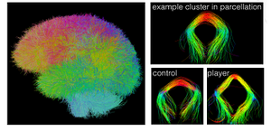
Figure 4: White matter segmentation using clustering for traumatic brain injury. Results from clustering the result of whole brain white matter tractography. Left: 650 color-coded clusters with a distinct color for each segmented tract segment. Right: Example of one selected cluster with the corresponding tract from the control group and the player group. By O'Donnell et al.

CF6

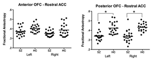
Figure 5: Using abnormal white matter connections between medial frontal regions to predict symptoms in patients with first episode schizophrenia. Scatter plots of mean fractional anisotropy for first episode schizophrenia and the healthy control group. Abbreviations: OFC = orbitofrontal cortex; ACC = anterior cingulate cortex; SZ = schizophrenia group; HC = healthy control group. *p < .001. ]

CF7

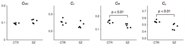
Figure 6: New microscopic measures derived from QTI applied to schizophrenia population. Comparison of normalized measures in schizophrenia patients (SZ) and healthy controls (CTR). The CMD, CM and Cμ averaged across the white matter were all significantly reduced in the schizophrenia group. Changes in Cc were not found to be significant between the groups. Significance was tested using the Wilcoxon rank-sum U-test. ]


PROGRESS UNDER AIM 1: DEVELOPMENT OF MICROSTRUCTURAL IMAGING BIOMARKERS

Dr. Westin’s team reported progress in four projects designed to further the goals of aim 1.

Q-space trajectory imaging for multidimensional diffusion MRI of the human brain (FIGURE 1). The team has developed a framework called q-space trajectory imaging (QTI), which uses gradient waveforms to probe trajectories in q-space. In contrast to single diffusion encoding (SDE) sequences that probe a single point in q-space, a single QTI measurement encodes multiple diffusion directions (Figure 1). The team has shown that measuring a family of trajectories introduces higher-order (multi-dimensional) correlations, which permit separate quantification of microstructural properties that are intrinsically entangled in traditional SDE. Within this framework the microstructural properties, mathematically expressed in terms of variability in size, shape, and orientation, are extracted from a diffusion tensor distribution model representing a mixture of distinct neuronal tissue micro-environments, such as neurites, cellular domains, and extracellular spaces [Westin, 2016].

Characterizing magnetic resonance signal decay using the path integral approach. The influence of Gaussian diffusion on the magnetic resonance signal is determined by the apparent diffusion coefficient (ADC) and tensor of the diffusing fluid as well as the gradient waveform applied to sensitize the signal to diffusion. Estimations of the ADC and the diffusion tensor from diffusion-weighted acquisitions necessitate computations of, respectively, the b-value and b-matrix associated with the employed pulse sequence. The team has established the relationship between these quantities and the gradient waveform by expressing the problem as a path integral, which is then explicitly evaluated. Further, the team has shown that these important quantities can be conveniently computed for any gradient waveform using a simple algorithm that requires a few lines of code. With this representation, the new technique complements the multiple correlation function method commonly used to compute the effects of restricted diffusion. It also provides a consistent and convenient framework for studies that aim to infer the microstructural features. To understand the meaning of MR signals, it is important, theoretically, to relate the applied waveform to an expected MR signal for a given structure or geometry. This has been studied extensively for single diffusion encoding (SDE). During the past year the team began this analysis for QTI by presenting the initial steps towards a general analytical framework with which one can derive explicit relationships for MR signal decay for a general time-dependent gradient waveform. Unlike earlier derivations, this one is based on path integrals and is more intuitive as it is based solely on probabilistic notions rather than the Bloch-Torrey equation. The team plans to extend this framework to develop efficient numerical methods for Gaussian and non-Gaussian diffusion models.

Constrained optimization of gradient waveforms for generalized diffusion encoding (FIGURE 2). The conventional diffusion encoding sequence, i.e., the single pulsed field gradient (PFG), has recently been challenged as more general gradient waveforms have been introduced, such as q-space trajectory imaging (QTI), which generalizes the scalar b-value to a tensor valued entity. To design gradient waveforms with specific features while taking full advantage of clinical scanner capabilities, it is imperative to respect the constraints imposed by the hardware while maximizing the diffusion encoding strength. The team has provided a tool that achieves this goal by solving a constrained optimization problem to accommodate constraints on maximum gradient amplitude, slew rate, coil heating, and positioning of radiofrequency pulses. The efficacy and flexibility of this method is demonstrated both experimentally and by comparison with previous work on optimization of isotropic diffusion sequences. Q-space trajectory imaging enables diffusion encoding with a general measurement tensor B. Although the “b-matrix” concept is well established and can be found in standard textbooks on diffusion NMR and MRI, the characterization of the b-matrix using double-PFG and more general gradient waveform diffusion MRI is novel and different. The team showed that it is possible to perform diffusion encoding imaging of the human brain (FIGURE 2) with arbitrary q-space trajectories while maintaining good SNR to generalize the concept that b-values can enable new types of measurements not available with single-PFG.

Sampling strategies and compressed sensing for high spatial resolution dMRI (FIGURES 3A, 3B). Compressed sensing (CS) is used in signal processing to reconstruct a signal from very few measurements or samples. CS has received significant attention in the MRI community for its ability to reconstruct an image with fewer data points, thereby reducing scan time. During the past year the team developed a novel sampling and reconstruction scheme for obtaining high spatial resolution dMRI images using multiple low resolution images that effectively reduces acquisition time while improving signal-to-noise (SNR) ratio. The proposed method, called compressed-sensing super-resolution reconstruction, uses multiple overlapping thick-slice dMRI volumes that are under-sampled in q-space to reconstruct diffusion signals with complex orientations. The proposed method combines the twin concepts of compressed sensing and super-resolution to model the diffusion signal (at a given b-value) using spherical ridgelets with total-variation regularization to account for signal correlation in neighboring voxels. The performance of the proposed method is quantitatively evaluated on several in vivo human data sets. The experimental results demonstrate that the proposed method can be used for reconstructing sub-millimeter super-resolution dMRI data with very good data fidelity in a clinically feasible acquisition time. 
 

PROGRESS UNDER AIM 2: DEVELOPMENT OF PERSONALIZED ANATOMICAL ANALYSIS METHODS

Dr. Westin’s team further refined their white matter tractography clustering method to permit automated measurement of white matter regions in subjects with traumatic brain injury.

White matter segmentation using clustering for traumatic brain injury (FIGURE 4). The team continued to develop a white matter atlas creation method for the purpose of studying subjects with traumatic brain injury (TBI). The method learns a model of the common white matter structures present in a group of subjects. Based on group spectral clustering of tractography, the method discovers structures corresponding to expected white matter anatomy such as the corpus callosum, uncinate fasciculus, cingulum bundles, arcuate fasciculus, and corona radiata. The white matter clusters are augmented with expert anatomical labels and stored in a new type of atlas called a high-dimensional white matter atlas. The automatic tractography segmentation identifies corresponding white matter regions across hemispheres and across subjects, thus enabling group comparison of white matter anatomy. This preliminary study analyzed the white matter structures of 63 retired National Football League (NFL) players vs 26 healthy age-matched (ages 45-69) controls. The clustering method enabled automated measurement of 650 white matter regions in each subject, with 7% of the clusters showing lower fractional anisotropy (FA) in the player group. 
 

PROGRESS UNDER AIM 3: APPLICATION TO TBI: POPULATION STATISTICS AND PATIENT-SPECIFIC DIAGNOSIS

Dr. Westin’s team developed new QTI analysis methods and applied them to population studies in schizophrenia.

Application of analysis methods to population studies in schizophrenia (FIGURE 5). The team studied abnormal white matter connections between medial frontal regions to determine their value in predicting symptoms in patients with first episode schizophrenia. Clinically, a majority of patients with early stages of schizophrenia, a population frequently referred to as first episode schizophrenia (FESZ), demonstrate positive symptoms such as hallucinations and delusions. Negative symptoms also exist in the early stage of the illness, although they are observed less frequently in FESZ than in chronic schizophrenia populations. It has been suggested that the medial orbitofrontal cortex (mOFC) and rostral part of anterior cingulate cortex (rACC) are involved in the neural network of salience and emotional processing and associated with specific clinical symptoms of schizophrenia. Considering the schizophrenia dysconnectivity hypothesis, the connectivity abnormalities between mOFC and rACC might be associated with clinical characteristics in first episode schizophrenia patients (Figure 5). Diffusion tensor imaging (DTI) is a promising method for characterizing microstructural changes or differences with neuropathology and treatment. Diffusion MRI (dMRI) encodes information on translational displacements of water on the micrometer scale. Thus, the dMRI signal is an excellent probe for microstructural geometries in tissue such as the human brain. In dMRI, each millimeter-scale measurement contains an aggregate of information from a multitude of microscopic environments (microenvironments). The diffusion tensor characterizes the magnitude, anisotropy, and orientation of the diffusion tensor to reveal the relationships between DTI measures and white matter pathologic features (ischemia, myelination, axonal damage, inflammation, and edema). FA is highly sensitive to microstrutural changes, but not very specific as to the orientation of the change. The main findings of this study include a reduction of FA and significant increases in radial diffusivity (RD) and Trace (magnitude) without significant changes in axial diffusivity (AD) in white matter fibers connecting the mOFC and the rACC, suggesting that dismyelination might affect normal connectivity in this region in patients with FESZ. In addition, FA reduction in the fibers connecting the posterior mOFC and rACC was related to the severity of delusions and emotion-related negative symptoms in FESZ, implying an association between posterior mOFC- rACC disconnectivity and the psychopathology of early stage of schizophrenia.

The team also derived new microscopic measures based on q-trajectory imaging (QTI) and applied them to a schizophrenia population (FIGURE 6). Parameter maps derived from QTI were compared between patients with chronic schizophrenia and healthy controls. Nine of the 14 parameters investigated showed differences between groups. The ability to measure and model the distribution of diffusion tensors, rather than a quantity that has already been averaged within a voxel, has the potential to provide a powerful paradigm for studying complex tissue architecture. Following a new approach, the team separated the bulk and shear variances into measures that are more intuitively meaningful, with the goal of separating size, shape, and orientation coherence. The measures were normalized and ranged between 0 and 1. Normalizing the bulk variance (VMD) yields a natural size variation parameter, CMD. Normalizing the shear variance is more challenging because it is affected by both the shape of the microenvironment and its orientation coherence (or dispersion). Using this approach, the team derived a measure of shape, Cμ, related to μFA and a separate measure of microscopic orientation coherence, Cc, related to previously presented order parameters (Figure 6). As expected, the schizophrenia patients showed elevated mean diffusivity (MD) in the white matter. Interestingly, this increase was matched by an increase in VMD. The increase cannot be explained by a homogeneous increase in the local mean diffusivity, rather it can be explained by an increase in the fraction of free water. This finding suggests that increased extracellular water, e.g., due to chronic neuroinflammatory processes or atrophy, is the primary mechanism underlying white matter diffusion changes in this cohort of patients with chronic schizophrenia. However, while reduced Cμ was also observed, Cc in the schizophrenia patients was unaltered, which could indicate axonal degradation at a microscopic level. Further studies with larger sample sizes are needed to confirm these findings. The current results, obtained by averaging throughout the entire white matter, do not lend themselves to the precise interpretation of the newly proposed measures in comparison with more traditional SDE measures. It may be more meaningful to study localized changes in both the white matter and the gray matter, and then to assess these results with analogy to simple prototype distributions.

Publications

  • Keijzer HM, Duering M, Pasternak O, Meijer FJA, Verhulst MMLH, Tonino BAR, Blans MJ, Hoedemaekers CWE, Klijn CJM, Hofmeijer J. Free Water Corrected Diffusion Tensor Imaging Discriminates Between Good and Poor Outcomes of Comatose Patients After Cardiac Arrest. Eur Radiol. 2023;33(3):2139–48.

    OBJECTIVES: Approximately 50% of comatose patients after cardiac arrest never regain consciousness. Cerebral ischaemia may lead to cytotoxic and/or vasogenic oedema, which can be detected by diffusion tensor imaging (DTI). Here, we evaluate the potential value of free water corrected mean diffusivity (MD) and fractional anisotropy (FA) based on DTI, for the prediction of neurological recovery of comatose patients after cardiac arrest. METHODS: A total of 50 patients after cardiac arrest were included in this prospective cohort study in two Dutch hospitals. DTI was obtained 2-4 days after cardiac arrest. Outcome was assessed at 6 months, dichotomised as poor (cerebral performance category 3-5; n = 20) or good (n = 30) neurological outcome. We calculated the whole brain mean MD and FA and compared between patients with good and poor outcomes. In addition, we compared a preliminary prediction model based on clinical parameters with or without the addition of MD and FA. RESULTS: We found significant differences between patients with good and poor outcome of mean MD (good: 726 [702-740] × 10-6 mm2/s vs. poor: 663 [575-736] × 10-6 mm2/s; p = 0.01) and mean FA (0.30 ± 0.03 vs. 0.28 ± 0.03; p = 0.03). An exploratory prediction model combining clinical parameters, MD and FA increased the sensitivity for reliable prediction of poor outcome from 60 to 85%, compared to the model containing clinical parameters only, but confidence intervals are overlapping. CONCLUSIONS: Free water-corrected MD and FA discriminate between patients with good and poor outcomes after cardiac arrest and hold the potential to add to multimodal outcome prediction. KEY POINTS: • Whole brain mean MD and FA differ between patients with good and poor outcome after cardiac arrest. • Free water-corrected MD can better discriminate between patients with good and poor outcome than uncorrected MD. • A combination of free water-corrected MD (sensitive to grey matter abnormalities) and FA (sensitive to white matter abnormalities) holds potential to add to the prediction of outcome.

  • Chakwizira A, Westin CF, Brabec J, Lasič S, Knutsson L, Szczepankiewicz F, Nilsson M. Diffusion MRI With Pulsed and Free Gradient Waveforms: Effects of Restricted Diffusion and Exchange. NMR Biomed. 2023;36(1):e4827.

    Monitoring time dependence with diffusion MRI yields observables sensitive to compartment sizes (restricted diffusion) and membrane permeability (water exchange). However, restricted diffusion and exchange have opposite effects on the diffusion-weighted signal, which can lead to errors in parameter estimates. In this work, we propose a signal representation that incorporates the effects of both restricted diffusion and exchange up to second order in b-value and is compatible with gradient waveforms of arbitrary shape. The representation features mappings from a gradient waveform to two scalars that separately control the sensitivity to restriction and exchange. We demonstrate that these scalars span a two-dimensional space that can be used to choose waveforms that selectively probe restricted diffusion or exchange, eliminating the correlation between the two phenomena. We found that waveforms with specific but unconventional shapes provide an advantage over conventional pulsed and oscillating gradient acquisitions. We also show that parametrization of waveforms into a two-dimensional space can be used to understand protocols from other approaches that probe restricted diffusion and exchange. For example, we found that the variation of mixing time in filter-exchange imaging corresponds to variation of our exchange-weighting scalar at a fixed value of the restriction-weighting scalar. The proposed signal representation was evaluated using Monte Carlo simulations in identical parallel cylinders with hexagonal and random packing as well as parallel cylinders with gamma-distributed radii. Results showed that the approach is sensitive to sizes in the interval 4-12 μm $$ \upmu \mathrmm $$ and exchange rates in the simulated range of 0 to 20 s - 1 $$ \mathrms^-1 $$ , but also that there is a sensitivity to the extracellular geometry. The presented theory constitutes a simple and intuitive description of how restricted diffusion and exchange influence the signal as well as a guide to protocol design capable of separating the two effects.

  • Brabec J, Friedjungová M, Vašata D, Englund E, Bengzon J, Knutsson L, Szczepankiewicz F, van Westen D, Sundgren PC, Nilsson M. Meningioma Microstructure Assessed by Diffusion MRI: An Investigation of the Source of Mean Diffusivity and Fractional Anisotropy by Quantitative Histology. NeuroImage Clin. 2023;37:103365.

    BACKGROUND: Mean diffusivity (MD) and fractional anisotropy (FA) from diffusion MRI (dMRI) have been associated with cell density and tissue anisotropy across tumors, but it is unknown whether these associations persist at the microscopic level.

    PURPOSE: To quantify the degree to which cell density and anisotropy, as determined from histology, account for the intra-tumor variability of MD and FA in meningioma tumors. Furthermore, to clarify whether other histological features account for additional intra-tumor variability of dMRI parameters.

    MATERIALS AND METHODS: We performed ex-vivo dMRI at 200 μm isotropic resolution and histological imaging of 16 excised meningioma tumor samples. Diffusion tensor imaging (DTI) was used to map MD and FA, as well as the in-plane FA (FAIP). Histology images were analyzed in terms of cell nuclei density (CD) and structure anisotropy (SA; obtained from structure tensor analysis) and were used separately in a regression analysis to predict MD and FAIP, respectively. A convolutional neural network (CNN) was also trained to predict the dMRI parameters from histology patches. The association between MRI and histology was analyzed in terms of out-of-sample (R2OS) on the intra-tumor level and within-sample R2 across tumors. Regions where the dMRI parameters were poorly predicted from histology were analyzed to identify features apart from CD and SA that could influence MD and FAIP, respectively.

    RESULTS: Cell density assessed by histology poorly explained intra-tumor variability of MD at the mesoscopic level (200 μm), as median R2OS = 0.04 (interquartile range 0.01-0.26). Structure anisotropy explained more of the variation in FAIP (median R2OS = 0.31, 0.20-0.42). Samples with low R2OS for FAIP exhibited low variations throughout the samples and thus low explainable variability, however, this was not the case for MD. Across tumors, CD and SA were clearly associated with MD (R2 = 0.60) and FAIP (R2 = 0.81), respectively. In 37% of the samples (6 out of 16), cell density did not explain intra-tumor variability of MD when compared to the degree explained by the CNN. Tumor vascularization, psammoma bodies, microcysts, and tissue cohesivity were associated with bias in MD prediction based solely on CD. Our results support that FAIP is high in the presence of elongated and aligned cell structures, but low otherwise.

    CONCLUSION: Cell density and structure anisotropy account for variability in MD and FAIP across tumors but cell density does not explain MD variations within the tumor, which means that low or high values of MD locally may not always reflect high or low tumor cell density. Features beyond cell density need to be considered when interpreting MD.

  • Zekelman LR, Zhang F, Makris N, He J, Chen Y, Xue T, Liera D, Drane DL, Rathi Y, Golby AJ, Donnell LJO. White Matter Association Tracts Underlying Language and Theory of Mind: An Investigation of 809 Brains from the Human Connectome Project. Neuroimage. 2022;246:118739.

    Language and theory of mind (ToM) are the cognitive capacities that allow for the successful interpretation and expression of meaning. While functional MRI investigations are able to consistently localize language and ToM to specific cortical regions, diffusion MRI investigations point to an inconsistent and sometimes overlapping set of white matter tracts associated with these two cognitive domains. To further examine the white matter tracts that may underlie these domains, we use a two-tensor tractography method to investigate the white matter microstructure of 809 participants from the Human Connectome Project. 20 association white matter tracts (10 in each hemisphere) are uniquely identified by leveraging a neuroanatomist-curated automated white matter tract atlas. The mean fractional anisotropy (FA), mean diffusivity (MD), and number of streamlines (NoS) are measured for each white matter tract. Performance on neuropsychological assessments of semantic memory (NIH Toolbox Picture Vocabulary Test, TPVT) and emotion perception (Penn Emotion Recognition Test, PERT) are used to measure critical subcomponents of the language and ToM networks, respectively. Regression models are constructed to examine how structural measurements of left and right white matter tracts influence performance across these two assessments. We find that semantic memory performance is influenced by the number of streamlines of the left superior longitudinal fasciculus III (SLF-III), and emotion perception performance is influenced by the number of streamlines of the right SLF-III. Additionally, we find that performance on both semantic memory & emotion perception is influenced by the FA of the left arcuate fasciculus (AF). The results point to multiple, overlapping white matter tracts that underlie the cognitive domains of language and ToM. Results are discussed in terms of hemispheric dominance and concordance with prior investigations.

  • Brabec J, Szczepankiewicz F, Lennartsson F, Englund E, Pebdani H, Bengzon J, Knutsson L, Westin CF, Sundgren PC, Nilsson M. Histogram Analysis of Tensor-Valued Diffusion MRI in Meningiomas: Relation to Consistency, Histological Grade and Type. Neuroimage Clin. 2022;33:102912.

    BACKGROUND: Preoperative radiological assessment of meningioma characteristics is of value for pre- and post-operative patient management, counselling, and surgical approach. PURPOSE: To investigate whether tensor-valued diffusion MRI can add to the preoperative prediction of meningioma consistency, grade and type. MATERIALS AND METHODS: 30 patients with intracranial meningiomas (22 WHO grade I, 8 WHO grade II) underwent MRI prior to surgery. Diffusion MRI was performed with linear and spherical b-tensors with b-values up to 2000 s/mm2. The data were used to estimate mean diffusivity (MD), fractional anisotropy (FA), mean kurtosis (MK) and its components-the anisotropic and isotropic kurtoses (MKA and MKI). Meningioma consistency was estimated for 16 patients during resection based on ultrasonic aspiration intensity, ease of resection with instrumentation or suction. Grade and type were determined by histopathological analysis. The relation between consistency, grade and type and dMRI parameters was analyzed inside the tumor ("whole-tumor") and within brain tissue in the immediate periphery outside the tumor ("rim") by histogram analysis. RESULTS: Lower 10th percentiles of MK and MKA in the whole-tumor were associated with firm consistency compared with pooled soft and variable consistency (n = 7 vs 9; U test, p = 0.02 for MKA 10 and p = 0.04 for MK10) and lower 10th percentile of MD with variable against soft and firm (n = 5 vs 11; U test, p = 0.02). Higher standard deviation of MKI in the rim was associated with lower grade (n = 22 vs 8; U test, p = 0.04) and in the MKI maps we observed elevated rim-like structure that could be associated with grade. Higher median MKA and lower median MKI distinguished psammomatous type from other pooled meningioma types (n = 5 vs 25; U test; p = 0.03 for MKA 50 and p = 0.03 and p = 0.04 for MKI 50). CONCLUSION: Parameters from tensor-valued dMRI can facilitate prediction of consistency, grade and type.

  • Ji Y, Hoge S, Gagoski B, Westin CF, Rathi Y, Ning L. Accelerating Joint Relaxation-Diffusion MRI by Integrating Time Division Multiplexing and Simultaneous Multi-Slice (TDM-SMS) Strategies. Magn Reson Med. 2022;87(6):2697–709.

    PURPOSE: To accelerate the acquisition of relaxation-diffusion imaging by integrating time-division multiplexing (TDM) with simultaneous multi-slice (SMS) for EPI and evaluate imaging quality and diffusion measures. METHODS: The time-division multiplexing (TDM) technique and SMS method were integrated to achieve a high slice-acceleration (e.g., 6×) factor for acquiring relaxation-diffusion MRI. Two variants of the sequence, referred to as TDM3e-SMS and TDM2s-SMS, were developed to simultaneously acquire slice groups with three distinct TEs and two slice groups with the same TE, respectively. Both sequences were evaluated on a 3T scanner with in vivo human brains and compared with standard single-band (SB) -EPI and SMS-EPI using diffusion measures and tractography results. RESULTS: Experimental results showed that the TDM3e-SMS sequence with total slice acceleration of 6 (multiplexing factor (MP) = 3 × multi-band factor (MB) = 2) provided similar image intensity and microstructure measures compared to standard SMS-EPI with MB = 2, and yielded less bias in intensity compared to standard SMS-EPI with MB = 4. The three sequences showed a similar positive correlation between TE and mean kurtosis (MK) and a negative correlation between TE and mean diffusivity (MD) in white matter. Multi-fiber tractography also shows consistency of results in TE-dependent measures between different sequences. The TDM2s-SMS sequence (MP = 2, MB = 2) also provided imaging measures similar to standard SMS-EPI sequences (MB = 2) for single-TE diffusion imaging. CONCLUSIONS: The TDM-SMS sequence can provide additional 2x to 3x acceleration to SMS without degrading imaging quality. With the significant reduction in scan time, TDM-SMS makes joint relaxation-diffusion MRI a feasible technique in neuroimaging research to investigate new markers of brain disorders.

  • Zhang F, Daducci A, He Y, Schiavi S, Seguin C, Smith RE, Yeh CH, Zhao T, Donnell LJO. Quantitative Mapping of the Brain’s Structural Connectivity Using Diffusion MRI Tractography: A Review. Neuroimage. 2022;249:118870.

    Diffusion magnetic resonance imaging (dMRI) tractography is an advanced imaging technique that enables in vivo reconstruction of the brain’s white matter connections at macro scale. It provides an important tool for quantitative mapping of the brain’s structural connectivity using measures of connectivity or tissue microstructure. Over the last two decades, the study of brain connectivity using dMRI tractography has played a prominent role in the neuroimaging research landscape. In this paper, we provide a high-level overview of how tractography is used to enable quantitative analysis of the brain’s structural connectivity in health and disease. We focus on two types of quantitative analyses of tractography, including: 1) tract-specific analysis that refers to research that is typically hypothesis-driven and studies particular anatomical fiber tracts, and 2) connectome-based analysis that refers to research that is more data-driven and generally studies the structural connectivity of the entire brain. We first provide a review of methodology involved in three main processing steps that are common across most approaches for quantitative analysis of tractography, including methods for tractography correction, segmentation and quantification. For each step, we aim to describe methodological choices, their popularity, and potential pros and cons. We then review studies that have used quantitative tractography approaches to study the brain’s white matter, focusing on applications in neurodevelopment, aging, neurological disorders, mental disorders, and neurosurgery. We conclude that, while there have been considerable advancements in methodological technologies and breadth of applications, there nevertheless remains no consensus about the "best" methodology in quantitative analysis of tractography, and researchers should remain cautious when interpreting results in research and clinical applications.

  • Seitz-Holland J, Seethaler M, Makris N, Rushmore J, Cho KIK, Rizzoni E, Vangel M, Sahin OS, Heller C, Pasternak O, Szczepankiewicz F, Westin CF, Lošák J, Ustohal L, Tomandl J, Vojtíšek L, Kudlička P, Jáni M, Woo W, Kašpárek T, Kikinis Z, Kubicki M. The Association of Matrix Metalloproteinase 9 (MMP9) With Hippocampal Volume in Schizophrenia: A Preliminary MRI Study. Neuropsychopharmacology. 2022;47(2):524–30.

    Matrix metalloproteinases 9 (MMP9) are enzymes involved in regulating neuroplasticity in the hippocampus. This, combined with evidence for disrupted hippocampal structure and function in schizophrenia, has prompted our current investigation into the relationship between MMP9 and hippocampal volumes in schizophrenia. 34 healthy individuals (mean age = 32.50, male = 21, female = 13) and 30 subjects with schizophrenia (mean age = 33.07, male = 19, female = 11) underwent a blood draw and T1-weighted magnetic resonance imaging. The hippocampus was automatically segmented utilizing FreeSurfer. MMP9 plasma levels were measured with ELISA. ANCOVAs were conducted to compare MMP9 plasma levels (corrected for age and sex) and hippocampal volumes between groups (corrected for age, sex, total intracranial volume). Spearman correlations were utilized to investigate the relationship between symptoms, medication, duration of illness, number of episodes, and MMP9 plasma levels in patients. Last, we explored the correlation between MMP9 levels and hippocampal volumes in patients and healthy individuals separately. Patients displayed higher MMP9 plasma levels than healthy individuals (F(1, 60) = 21.19, p < 0.0001). MMP9 levels correlated with negative symptoms in patients (R = 0.39, p = 0.035), but not with medication, duration of illness, or the number of episodes. Further, patients had smaller left (F(1,59) = 9.12, p = 0.0040) and right (F(1,59) = 6.49, p = 0.013) hippocampal volumes. Finally, left (R = -0.39, p = 0.034) and right (R = -0.37, p = 0.046) hippocampal volumes correlated negatively with MMP9 plasma levels in patients. We observe higher MMP9 plasma levels in SCZ, associated with lower hippocampal volumes, suggesting involvement of MMP9 in the pathology of SCZ. Future studies are needed to investigate how MMP9 influences the pathology of SCZ over the lifespan, whether the observed associations are specific for schizophrenia, and if a therapeutic modulation of MMP9 promotes neuroprotective effects in SCZ.

  • Zhang F, Cho KIK, Tang Y, Zhang T, Kelly S, Di Biase M, Xu L, Li H, Matcheri K, Whitfield-Gabrieli S, Niznikiewicz M, Stone WS, Wang J, Shenton ME, Pasternak O. MK-Curve Improves Sensitivity to Identify White Matter Alterations in Clinical High Risk for Psychosis. Neuroimage. 2021;226:117564.

    Diffusion kurtosis imaging (DKI) is a diffusion MRI approach that enables the measurement of brain microstructural properties, reflecting molecular restrictions and tissue heterogeneity. DKI parameters such as mean kurtosis (MK) provide additional subtle information to that provided by popular diffusion tensor imaging (DTI) parameters, and thus have been considered useful to detect white matter abnormalities, especially in populations that are not expected to show severe brain pathologies. However, DKI parameters often yield artifactual output values that are outside of the biologically plausible range, which diminish sensitivity to identify true microstructural changes. Recently we have proposed the mean-kurtosis-curve (MK-Curve) method to correct voxels with implausible DKI parameters, and demonstrated its improved performance against other approaches that correct artifacts in DKI. In this work, we aimed to evaluate the utility of the MK-Curve method to improve the identification of white matter abnormalities in group comparisons. To do so, we compared group differences, with and without the MK-Curve correction, between 115 individuals at clinical high risk for psychosis (CHR) and 93 healthy controls (HCs). We also compared the correlation of the corrected and uncorrected DKI parameters with clinical characteristics. Following the MK-curve correction, the group differences had larger effect sizes and higher statistical significance (i.e., lower p-values), demonstrating increased sensitivity to detect group differences, in particular in MK. Furthermore, the MK-curve-corrected DKI parameters displayed stronger correlations with clinical variables in CHR individuals, demonstrating the clinical relevance of the corrected parameters. Overall, following the MK-curve correction our analyses found widespread lower MK in CHR that overlapped with lower fractional anisotropy (FA), and both measures were significantly correlated with a decline in functioning and with more severe symptoms. These observations further characterize white matter alterations in the CHR stage, demonstrating that MK and FA abnormalities are widespread, and mostly overlap. The improvement in group differences and stronger correlation with clinical variables suggest that applying MK-curve would be beneficial for the detection and characterization of subtle group differences in other experiments as well.

  • Ramos-Llordén G, Vegas-Sánchez-Ferrero G, Liao C, Westin CF, Setsompop K, Rathi Y. SNR-Enhanced Diffusion MRI With Structure-Preserving Low-Rank Denoising in Reproducing Kernel Hilbert Spaces. Magn Reson Med. 2021;86(3):1614–32.

    PURPOSE: To introduce, develop, and evaluate a novel denoising technique for diffusion MRI that leverages nonlinear redundancy in the data to boost the SNR while preserving signal information. METHODS: We exploit nonlinear redundancy of the dMRI data by means of kernel principal component analysis (KPCA), a nonlinear generalization of PCA to reproducing kernel Hilbert spaces. By mapping the signal to a high-dimensional space, a higher level of redundant information is exploited, thereby enabling better denoising than linear PCA. We implement KPCA with a Gaussian kernel, with parameters automatically selected from knowledge of the noise statistics, and validate it on realistic Monte Carlo simulations as well as with in vivo human brain submillimeter and low-resolution dMRI data. We also demonstrate KPCA denoising on multi-coil dMRI data. RESULTS: SNR improvements up to 2.7 were obtained in real in vivo datasets denoised with KPCA, in comparison to SNR gains of up to 1.8 using a linear PCA denoising technique called Marchenko-Pastur PCA (MPPCA). Compared to gold-standard dataset references created from averaged data, we showed that lower normalized root mean squared error was achieved with KPCA compared to MPPCA. Statistical analysis of residuals shows that anatomical information is preserved and only noise is removed. Improvements in the estimation of diffusion model parameters such as fractional anisotropy, mean diffusivity, and fiber orientation distribution functions were also demonstrated. CONCLUSION: Nonlinear redundancy of the dMRI signal can be exploited with KPCA, which allows superior noise reduction/SNR improvements than the MPPCA method, without loss of signal information.

  • Zhang F, Breger A, Cho KIK, Ning L, Westin CF, Donnell LJO, Pasternak O. Deep Learning Based Segmentation of Brain Tissue from Diffusion MRI. Neuroimage. 2021;233:117934.

    Segmentation of brain tissue types from diffusion MRI (dMRI) is an important task, required for quantification of brain microstructure and for improving tractography. Current dMRI segmentation is mostly based on anatomical MRI (e.g., T1- and T2-weighted) segmentation that is registered to the dMRI space. However, such inter-modality registration is challenging due to more image distortions and lower image resolution in dMRI as compared with anatomical MRI. In this study, we present a deep learning method for diffusion MRI segmentation, which we refer to as DDSeg. Our proposed method learns tissue segmentation from high-quality imaging data from the Human Connectome Project (HCP), where registration of anatomical MRI to dMRI is more precise. The method is then able to predict a tissue segmentation directly from new dMRI data, including data collected with different acquisition protocols, without requiring anatomical data and inter-modality registration. We train a convolutional neural network (CNN) to learn a tissue segmentation model using a novel augmented target loss function designed to improve accuracy in regions of tissue boundary. To further improve accuracy, our method adds diffusion kurtosis imaging (DKI) parameters that characterize non-Gaussian water molecule diffusion to the conventional diffusion tensor imaging parameters. The DKI parameters are calculated from the recently proposed mean-kurtosis-curve method that corrects implausible DKI parameter values and provides additional features that discriminate between tissue types. We demonstrate high tissue segmentation accuracy on HCP data, and also when applying the HCP-trained model on dMRI data from other acquisitions with lower resolution and fewer gradient directions.

  • Szczepankiewicz F, Westin CF, Nilsson M. Gradient Waveform Design for Tensor-valued Encoding in Diffusion MRI. J Neurosci Methods. 2021;348:109007.

    Diffusion encoding along multiple spatial directions per signal acquisition can be described in terms of a b-tensor. The benefit of tensor-valued diffusion encoding is that it unlocks the ’shape of the b-tensor’ as a new encoding dimension. By modulating the b-tensor shape, we can control the sensitivity to microscopic diffusion anisotropy which can be used as a contrast mechanism; a feature that is inaccessible by conventional diffusion encoding. Since imaging methods based on tensor-valued diffusion encoding are finding an increasing number of applications we are prompted to highlight the challenge of designing the optimal gradient waveforms for any given application. In this review, we first establish the basic design objectives in creating field gradient waveforms for tensor-valued diffusion MRI. We also survey additional design considerations related to limitations imposed by hardware and physiology, potential confounding effects that cannot be captured by the b-tensor, and artifacts related to the diffusion encoding waveform. Throughout, we discuss the expected compromises and tradeoffs with an aim to establish a more complete understanding of gradient waveform design and its impact on accurate measurements and interpretations of data.

  • Maziero MP, Seitz-Holland J, Cho KIK, Goldenberg JE, Tanamatis TW, Diniz JB, Cappi C, de Mathis MA, Otaduy MCG, Martin M da GM, da Silva R de MF, Shavitt RG, Batistuzzo MC, Lopes AC, Miguel EC, Pasternak O, Hoexter MQ. Cellular and Extracellular White Matter Abnormalities in Obsessive-Compulsive Disorder: A Diffusion MRI Study. Biol Psychiatry Cogn Neurosci Neuroimaging. 2021;6(10):983–91.

    BACKGROUND: While previous studies have implicated white matter (WM) as a core pathology of Obsessive-Compulsive Disorder (OCD), the underlying neurobiological processes remain elusive. This study utilizes free-water imaging derived from diffusion MRI to identify cellular and extracellular WM abnormalities in patients with OCD compared to controls (Cs). Next, we investigate the association between diffusion measures, and clinical variables in patients. METHODS: We collected diffusion-weighted MRI and clinical data from eighty-three patients with OCD (56 females/27 males, age=37.7 ± 10.6) and 52 Cs (27 females/25 males, age=32.8 ± 11.5). Fractional anisotropy (FA), fractional anisotropy of cellular tissue (FAT), and extracellular free-water (FW) maps were extracted and compared between patients and Cs using tract-based spatial statistics, and voxel-wise comparison in FSL’s Randomise. Next, we correlated these WM measures with clinical variables (age-of-onset and symptom severity) and compared them between patients with and without comorbidities and patients with and without psychiatric medication. RESULTS: Patients with OCD demonstrated lower FA (43.4% of the WM skeleton), FAт (31% of the WM skeleton), and higher FW (22.5% of the WM skeleton) compared to Cs. We did not observe significant correlations between diffusion measures and clinical variables. Comorbidities and medication status did not influence diffusion measures. CONCLUSIONS: Our findings of widespread FA, FAт, and FW abnormalities suggest that OCD is associated with both microstructural cellular and extracellular abnormalities beyond the cortico-striato-thalamo-cortical circuits. Future multimodal longitudinal studies are needed to understand better the influence of essential clinical variables across the illness trajectory.

  • He J, Zhang F, Xie G, Yao S, Feng Y, Bastos DCA, Rathi Y, Makris N, Kikinis R, Golby AJ, Donnell LJO. Comparison of Multiple Tractography Methods for Reconstruction of the Retinogeniculate Visual Pathway Using Diffusion MRI. Hum Brain Mapp. 2021;42(12):3887–904.

    The retinogeniculate visual pathway (RGVP) conveys visual information from the retina to the lateral geniculate nucleus. The RGVP has four subdivisions, including two decussating and two nondecussating pathways that cannot be identified on conventional structural magnetic resonance imaging (MRI). Diffusion MRI tractography has the potential to trace these subdivisions and is increasingly used to study the RGVP. However, it is not yet known which fiber tracking strategy is most suitable for RGVP reconstruction. In this study, four tractography methods are compared, including constrained spherical deconvolution (CSD) based probabilistic (iFOD1) and deterministic (SD-Stream) methods, and multi-fiber (UKF-2T) and single-fiber (UKF-1T) unscented Kalman filter (UKF) methods. Experiments use diffusion MRI data from 57 subjects in the Human Connectome Project. The RGVP is identified using regions of interest created by two clinical experts. Quantitative anatomical measurements and expert anatomical judgment are used to assess the advantages and limitations of the four tractography methods. Overall, we conclude that UKF-2T and iFOD1 produce the best RGVP reconstruction results. The iFOD1 method can better quantitatively estimate the percentage of decussating fibers, while the UKF-2T method produces reconstructed RGVPs that are judged to better correspond to the known anatomy and have the highest spatial overlap across subjects. Overall, we find that it is challenging for current tractography methods to both accurately track RGVP fibers that correspond to known anatomy and produce an approximately correct percentage of decussating fibers. We suggest that future algorithm development for RGVP tractography should take consideration of both of these two points.

  • Guder S, Pasternak O, Gerloff C, Schulz R. Strengthened Structure-Function Relationships of the Corticospinal Tract by Free Water Correction After Stroke. Brain Commun. 2021;3(2):fcab034.

    The corticospinal tract is the most intensively investigated tract of the human motor system in stroke rehabilitative research. Diffusion-tensor-imaging gives insights into its microstructure, and transcranial magnetic stimulation assesses its excitability. Previous data on the interrelationship between both measures are contradictory. Correlative or predictive models which associate them with motor outcome are incomplete. Free water correction has been developed to enhance diffusion-tensor-imaging by eliminating partial volume with extracellular water, which could improve capturing stroke-related microstructural alterations, thereby also improving structure-function relationships in clinical cohorts. In the present cross-sectional study, data of 18 chronic stroke patients and 17 healthy controls, taken from a previous study on cortico-cerebellar motor tracts, were re-analysed: The data included diffusion-tensor-imaging data quantifying corticospinal tract microstructure with and without free water correction, transcranial magnetic stimulation data assessing recruitment curve properties of motor evoked potentials and detailed clinical data. Linear regression modelling was used to interrelate corticospinal tract microstructure, recruitment curves properties and clinical scores. The main finding of the present study was that free water correction substantially strengthens structure-function associations in stroke patients: Specifically, our data evidenced a significant association between fractional anisotropy of the ipsilesional corticospinal tract and its excitability ( = 0.001, adj. = 0.54), with free water correction explaining additional 20% in recruitment curve variability. For clinical scores, only free water correction leads to the reliable detection of significant correlations between ipsilesional corticospinal tract fractional anisotropy and residual grip ( = 0.001, adj. = 0.70) and pinch force ( < 0.001, adj. = 0.72). Finally, multimodal models can be improved by free water correction as well. This study evidences that corticospinal tract microstructure directly relates to its excitability in stroke patients. It also shows that unexplained variance in motor outcome is considerably reduced by free water correction arguing that it might serve as a powerful tool to improve existing models of structure-function associations and potentially also outcome prediction after stroke.

  • Langbein BJ, Szczepankiewicz F, Westin CF, Bay C, Maier SE, Kibel AS, Tempany CM, Fennessy FM. A Pilot Study of Multidimensional Diffusion MRI for Assessment of Tissue Heterogeneity in Prostate Cancer. Invest Radiol. 2021;56(12):845–53.

    OBJECTIVES: The objectives of this exploratory study were to investigate the feasibility of multidimensional diffusion magnetic resonance imaging (MddMRI) in assessing diffusion heterogeneity at both a macroscopic and microscopic level in prostate cancer (PCa). MATERIALS AND METHODS: Informed consent was obtained from 46 subjects who underwent 3.0-T prostate multiparametric MRI, complemented with a prototype spin echo-based MddMRI sequence in this institutional review board-approved study. Prostate cancer tumors and comparative normal tissue from each patient were contoured on both apparent diffusion coefficient and MddMRI-derived mean diffusivity (MD) maps (from which microscopic diffusion heterogeneity [MKi] and microscopic diffusion anisotropy were derived) using 3D Slicer. The discriminative ability of MddMRI-derived parameters to differentiate PCa from normal tissue was determined using the Friedman test. To determine if tumor diffusion heterogeneity is similar on macroscopic and microscopic scales, the linear association between SD of MD and mean MKi was estimated using robust regression (bisquare weighting). Hypothesis testing was 2 tailed; P values less than 0.05 were considered statistically significant. RESULTS: All MddMRI-derived parameters could distinguish tumor from normal tissue in the fixed-effects analysis (P < 0.0001). Tumor MKi was higher (P < 0.05) compared with normal tissue (median, 0.40; interquartile range, 0.29-0.52 vs 0.20-0.18; 0.25), as was tumor microscopic diffusion anisotropy (0.55; 0.36-0.81 vs 0.20-0.15; 0.28). The MKi could not be predicted (no significant association) by SD of MD. There was a significant correlation between tumor volume and SD of MD (R2 = 0.50, slope = 0.008 μm2/ms per millimeter, P < 0.001) but not between tumor volume and MKi. CONCLUSIONS: This explorative study demonstrates that MddMRI provides novel information on MKi and microscopic anisotropy, which differ from measures at the macroscopic level. MddMRI has the potential to characterize tumor tissue heterogeneity at different spatial scales.

  • Behjat H, Aganj I, Abramian D, Eklund A, Westin CF. Characterization of Spatial Dynamics of Fmri Data in White Matter Using Diffusion-Informed White Matter Harmonics. Proc IEEE Int Symp Biomed Imaging. 2021;2021:1586–90.

    In this work, we leverage the Laplacian eigenbasis of voxel-wise white matter (WM) graphs derived from diffusion-weighted MRI data, dubbed WM harmonics, to characterize the spatial structure of WM fMRI data. Our motivation for such a characterization is based on studies that show WM fMRI data exhibit a spatial correlational anisotropy that coincides with underlying fiber patterns. By quantifying the energy content of WM fMRI data associated with subsets of WM harmonics across multiple spectral bands, we show that the data exhibits notable subtle spatial modulations under functional load that are not manifested during rest. WM harmonics provide a novel means to study the spatial dynamics of WM fMRI data, in such way that the analysis is informed by the underlying anatomical structure.

  • Slator PJ, Palombo M, Miller KL, Westin CF, Laun F, Kim D, Haldar JP, Benjamini D, Lemberskiy G, Martins JP de A, Hutter J. Combined Diffusion-Relaxometry Microstructure Imaging: Current Status and Future Prospects. Magn Reson Med. 2021;86(6):2987–3011.

    Microstructure imaging seeks to noninvasively measure and map microscopic tissue features by pairing mathematical modeling with tailored MRI protocols. This article reviews an emerging paradigm that has the potential to provide a more detailed assessment of tissue microstructure-combined diffusion-relaxometry imaging. Combined diffusion-relaxometry acquisitions vary multiple MR contrast encodings-such as b-value, gradient direction, inversion time, and echo time-in a multidimensional acquisition space. When paired with suitable analysis techniques, this enables quantification of correlations and coupling between multiple MR parameters-such as diffusivity, T 1 , T 2 , and T 2 * . This opens the possibility of disentangling multiple tissue compartments (within voxels) that are indistinguishable with single-contrast scans, enabling a new generation of microstructural maps with improved biological sensitivity and specificity.

  • Di Biase MA, Cetin-Karayumak S, Lyall AE, Zalesky A, Cho KIK, Zhang F, Kubicki M, Rathi Y, Lyons MG, Bouix S, Billah T, Anticevic A, Schleifer C, Adkinson BD, Ji JL, Tamayo Z, Addington J, Bearden CE, Cornblatt BA, Keshavan MS, Mathalon DH, McGlashan TH, Perkins DO, Cadenhead KS, Tsuang MT, Woods SW, Stone WS, Shenton ME, Cannon TD, Pasternak O. White Matter Changes in Psychosis Risk Relate to Development and Are Not Impacted by the Transition to Psychosis. Mol Psychiatry. 2021;26(11):6833–44.

    Subtle alterations in white matter microstructure are observed in youth at clinical high risk (CHR) for psychosis. However, the timing of these changes and their relationships to the emergence of psychosis remain unclear. Here, we track the evolution of white matter abnormalities in a large, longitudinal cohort of CHR individuals comprising the North American Prodrome Longitudinal Study (NAPLS-3). Multi-shell diffusion magnetic resonance imaging data were collected across multiple timepoints (1-5 over 1 year) in 286 subjects (aged 12-32 years): 25 CHR individuals who transitioned to psychosis (CHR-P; 61 scans), 205 CHR subjects with unknown transition outcome after the 1-year follow-up period (CHR-U; 596 scans), and 56 healthy controls (195 scans). Linear mixed effects models were fitted to infer the impact of age and illness-onset on variation in the fractional anisotropy of cellular tissue (FAT) and the volume fraction of extracellular free water (FW). Baseline measures of white matter microstructure did not differentiate between HC, CHR-U and CHR-P individuals. However, age trajectories differed between the three groups in line with a developmental effect: CHR-P and CHR-U groups displayed higher FAT in adolescence, and 4% lower FAT by 30 years of age compared to controls. Furthermore, older CHR-P subjects (20+ years) displayed 4% higher FW in the forceps major (p < 0.05). Prospective analysis in CHR-P did not reveal a significant impact of illness onset on regional FAT or FW, suggesting that transition to psychosis is not marked by dramatic change in white matter microstructure. Instead, clinical high risk for psychosis-regardless of transition outcome-is characterized by subtle age-related white matter changes that occur in tandem with development.

  • Behjat H, Westin CF, Aganj I. Cortical Surface-Informed Volumetric Spatial Smoothing of fMRI Data via Graph Signal Processing. Annu Int Conf IEEE Eng Med Biol Soc. 2021;2021:3804–8.

    Conventionally, as a preprocessing step, functional MRI (fMRI) data are spatially smoothed before further analysis, be it for activation mapping on task-based fMRI or functional connectivity analysis on resting-state fMRI data. When images are smoothed volumetrically, however, isotropic Gaussian kernels are generally used, which do not adapt to the underlying brain structure. Alternatively, cortical surface smoothing procedures provide the benefit of adapting the smoothing process to the underlying morphology, but require projecting volumetric data on to the surface. In this paper, leveraging principles from graph signal processing, we propose a volumetric spatial smoothing method that takes advantage of the gray-white and pial cortical surfaces, and as such, adapts the filtering process to the underlying morphological details at each point in the cortex.